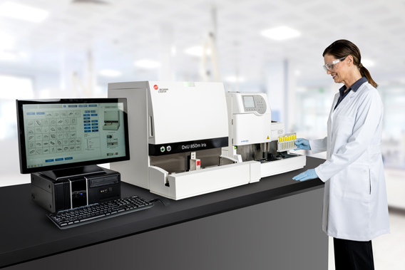

Routine microscopic analyses can provide better detection as to the quantity and morphology of RBCs which can further assist in identifying the pathogenic source and cause. Hematuria is normally categorized as glomerular, renal or urologic. However, this methodology is inherently suspect to interference by various factors that include medications and elevated urine concentrations of protein and ascorbic acid. Urine chemistry dipsticks detect for blood by measuring peroxidase activity of red blood cells (RBCs). This misdiagnosis is rather concerning as the presence of blood in urine is clinically significant, especially for males. Their study reported more than 25 percent of patients were misdiagnosed for microhematuria when relying solely on urine dipstick result. Brunzel and Barry highlighted the omission of the presence of blood and bacteria in normal samples.

1 The authors reported the negative predictive value (NPV) for their study using dipstick results and reflexing to microscopy was just 75 percent.

Such occurrences are by no means isolated events as investigations into the benefits and pitfalls of reflexing to microscopy have shown that omitting microscopy exams results in missed diagnosis, which of course causes repeated specimen collection and additional laboratory testing.Ī study authored by Nancy Brunzel and Diane Berry analyzing 709 routine specimens revealed reflexing to microscopy resulted in one in four patient samples being overlooked for pathological conditions. However, this practice may in fact have the opposite effect on laboratory efficiency and diminish patient care as urine samples with normal dipstick results have been later identified as containing pathogenic abnormalities. It’s reasonable to assume that excluding normal specimens from microscopic examinations would provide greater cost savings and faster turnaround-times (TAT). However, despite technical advances, even current users of automated urinalysis systems remain committed to the “old-school” practice of reflexing to microscopy as a continuous effort to improve productivity in the clinical laboratory.Īs more and more clinical and hospital labs adopt efficient and Lean principles as standard operating practice, more focus is placed on waste reduction while improving the quality of patient care. Advances in automated urine microscopy analyzers have significantly improved the workflow efficiencies and diagnostic proficiencies of modern urinalysis.
#Beckman coulter urinalysis manual
This is often common practice when manual microscopy is used to examine urine sediment, as it is one of the most tedious and time-consuming processes in the clinical laboratory. Reflex to microscopy refers to omitting normal specimens and only performing microscopic exams on urine specimens with abnormal dipstick results.

Microscopic analysis is a visual examination – qualitative and quantitative – of cellular and formed particles in the urine. Urine chemistry dipsticks test as many as 12 analytes that include blood (hemoglobin), protein, glucose and other urinary and metabolic biomarkers. In most, if not all labs, macroscopic analysis is performed at the same time as the chemical analysis. Total urinalysis comprises of macroscopic (visual observation), chemical (dipstick) and microscopic (urine sediment) analysis. Urinalysis provides valuable information, speaking not only to the well-being of the renal and urinary systems, but also to identifying metabolic, hemolytic and other pathological conditions.


 0 kommentar(er)
